With funding provided by the CIISB project, the NanoBio Core Facility has been equipped with new instruments that will help scientists from different disciplines to tackle their projects.
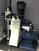
The laboratory's flagship is the large AFM microscope JPK NanoWizard 4XP installed on a Leica DMi8 optical microscope with a fluorescence module. Both microscopes can operate simultaneously in the so-called directoverlay mode, this combining AFM and optical microscopy abilities. Moreover, the new AFM microscope offers an extended operation range, compared to the previous model (NW 3) operated in our laboratory from 2014. The maximal range of operation is now extended to the 200x200x200 µm by hybrid stage, an essential factor for working with rugged samples such as bone sections or plant samples. The NanoWizard 4XP AFM microscope is not only an imaging tool, however, helps to map elastic properties of various samples with nanometer resolution. One of the main advantages is the ability to work in semi-physiological conditions.
Detailed an overall view of the JPK NanoWizard 4XP AFM microscope in the lab of the Nanobio Core Facility.
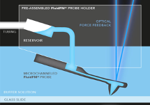
The new AFM device is equipped with a compelling module, the so-called FluidFM. This technology combines the precise nanomechanical indentation ability of the atomic force microscope with microfluidics. Microchannel inside the cantilever changes this tool into the nano-pipette, able to aspirate and/or deliver extremely low volumes. This feature can be used when injecting or removing small volumes from individual cells. Using stiffer cantilevers, the system can investigate cell adhesion on new types of implant materials.
Schematic view of the FluidFM module (left side), and the module in action (right side), when used for single-cell injection (schemes and images reprinted from https://www.blue-scientific.com).
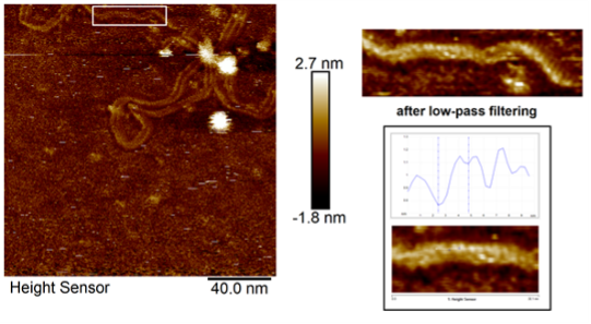
Keeping on the cutting-edge current AFM technology, the new generation of the MultiMode AFM microscope, version 8HR, was built for imaging with the maximum resolution that current commercial setups allow. This AFM setup will help the structural biologist image the biomolecules (DNA, proteins, molecular complexes) on a single molecular level.
New BioAFM Bruker MultiMode 8HR (left), maximum resolution illustrated on the right, where the double helix of ds plasmid structure is shown.
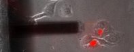
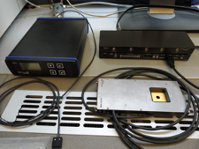
A multielectrode array (MEA) is a grid of tightly spaced microscopic electrodes embedded in the bottom of each well in a multi-well MEA plate. Cells, such as cardiomyocytes or neurons, electrically active, can be cultured over the electrodes creating a cohesive network. The functional behavior or electrical activity of this network can be recorded. These action potentials are recorded extracellularly and are known as field potentials. MEA device can be used to record spontaneous activity from hiPSC-derived neuronal cells upon differentiation and maturity, cell cultures for disease modeling, drug screening, or toxicology studies. Moreover, the MEA can be simultaneously connected with an AFM microscope, thus studying mechanoelectrical feedback of cardiac cells, tightly connected with some heart pathologies, such as catecholaminergic polymorphic ventricular tachycardia (CPVT).
Multielectrode array device MEA2100Lite from MultiChannel Instruments (left), cluster of iPSC cardiomyocytes grown on the chip containing a field of microelectrodes is shown on the right side (reprint from Biosensors and Bioelectronics 124–125 (2019) 129–135).
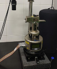
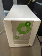
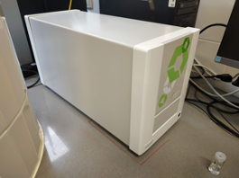
Upcon® is a first-of-a-kind complete solution utilizing upconversion technology. It offers a unique concept enabling high sensitivity and outstanding performance in life science applications, research, and diagnostics. The Upcon system includes easy-to-use and robust instruments, brightly luminescent upconverting nanoparticles, software, and technical support, bringing the great benefits of the technology to research laboratories and point-of-care applications. Labrox Upcon S pro reader newly installed in our lab is equipped with a 976 nm IR laser and suitable for a complex characterization of upconverting nanoparticles in absorbance and fluorescence (incl. time‑resolved and polarized) luminescence modes. The CFs users may bring their samples in 6-1536 well plates, both – top and bottom reading modes are available.
Labrox Upcon S pro reader newly installed in the Core Facility labs.